The lumbar spinal roots pass through the upper part of intervertebral foramen (Neuroforamen). However, they are fixed by ligaments which are the focus of our work. Each motor root travels anteromedial to the sensory portion. Both roots travel downward passing the frontal plane diagonally. In the intervertebral foramen they merge with these ligaments reaching the periphery. Physicians are aware of discrepancies between a lack of radiologic findings and syndromes, which are typical for spinal canal or foraminal stenosis.
We think that degenerative changes of extra- and intraforaminal ligaments or the surrounding soft tissue could be responsible for evoking such phenomena, similar to the known discogenic or stenotic diseases where a diagnosis is proven with a radiologic correlate. Thus, even without visible changes in radiological imaging these diseases, also known as "occult" stenosis, may occur. With support from Deutsche Arthrose-Hilfe (German Arthrosis Society) we investigate these reins-like ligaments, or "connectives", holding and tightening the spinal nerves through the lumbar intervertebral foramina.
(funded by Deutsche Arthrose-Hilfe)
The role of connective tissue in discogenic and stenotic syndromes
Hanno Steinke in Cooperation with Christoph Heyde (medical faculty), Dina Wiesbicki (orthopedics), Anna Völker (orthopedics), Jeanette Henkelmann (radiology), Charlotte Kulow (anatomy)
Funded by Deutsche Arthrose-Hilfe (DAH)
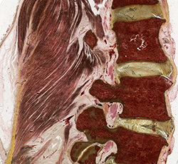
Our main hypothesis is that extra- and intraforaminal ligaments (EILs) are anatomical entities made of collagen. We assume that the healthy (waterbed-like) construction of an intervertebral disc depends on taut radial surrounding ligaments which are part of the intervertebral foramen. We are able to stain different collagen types close to the intervertebral foramen in plastinates (Fig. 1), confirmed by correlative Western Blotting.
In our second hypothesis we state that a pathological turnover of collagen types of these EILs close to the surface of the spinal nerve may cause spinal diseases. These are comparable to macroscopic degenerative changes. EILs are connected to caudal ligaments of the intervertebral disc and to the lower pedicle as well as to the ligamenta flava (Fig. 1). EILs also reach the ligaments of the facet joint. Due to these connections a minimal change in EILs may evoke symptoms, which are similar to pressing these ligaments by a thickened intervertebral disk (Nucleus pulposus prolapse, osteophytes) or by ligamenta flava hypertrophy, which is visible in MRI.
In our third hypothesis we postulate that a change of the collagen type II of the ligamenta flava leads to symptoms of a degenerative disease. Therefore even symptoms of facet joint diseases could be “invisble”. Degenerative changes seen in malexpression of collagen in the capsula of the facet joint may cause lower back pain without pathological findings in the MRI. This condition may happen as an early stage of degenerative disease.
Our hypotheses are based on surgical and postmortem human tissue samples.