You are here: Skip Navigation LinksCariology, Endodontology and Periodontology Research areas

WELCOME to our lab

​The research of our lab focusses on understanding the etiology of caries and the process of dental wear. To this end, we investigate dentin and enamel caries, the oral microbiome and wear surfaces. Furthermore, we measure the bonding between tooth, bone, dental materials and adjacent tissues applying 3D methods. We also develop mathematical models to simulate and link the measured 3D morphology to related disease patterns. The basic insights we gain this way, directly contribute to the development of novel treatment approaches in periodontal and endodontic dentistry.​

Periodontology, Cariology, Endodontology

​Dental caries is the most prevalent disease worldwide. With this in mind, we develop new approaches for caries diagnostics and monitoring, and we test existing approaches for validity and reliability. We analyze and quantify the oral microbiome to detect the amount of the cariogenic biofilm and to develop new therapeutic approaches. Furthermore, multidisciplinary dental care is an important research area to access oral health-related quality of life. Therefore, we pursue the approach of cross-sectional case studies in patients with chronical diseases (e. g. diabetes, severe heart failure, rheumatoid arthritis) to examine oral health, behavior and life quality. Moreover, we aim to develop concepts for endo- and periodontal treatments.​


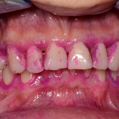
Intraoral view of stained dental biofilm, with old (violet) and new (red) plaque areas
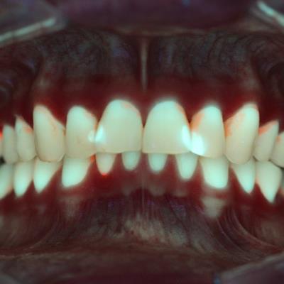
Red fluorescence indicates areas with bacterial metabolites (porphyrines) in the biofilm (QLF-D Biluminator)
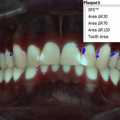

Blue areas indicate quantitative assessment of the fluorescence signal (QLF-D Biluminator)
Restorative and Adhesive Dentistry

Our goal is to develop efficient real-time assessments of adhesive-composite/ceramic restorations in frontal and cheek teeth to identify clinically relevant evaluation parameters. Thus, we conduct controlled clinical in-vitro as well as in-vivo trials to access the performance of adhesives in different application modes. Our expertise is the development and use of innovative quantitative 3D imaging techniques such as optical coherence tomography, µCT and 3D surface scanning. We visualize and measure failure of the composite structure and develop new clinical therapy concepts in prophylaxis.​


Optical coherence tomography using the Telesto II sensor (Thorlabs)

In-vivo composite filling (class V, canine) with prolonged gap at the dentin-composite-interface

µCT (SkyScan 1172-100-50 Bruker)

Composite filling (class II, premolar), µCT-cross section with diverse tooth and restoration structures

Chewing Efficiency and Tooth Wear

The research area of 3D wear analyses is evolving on all scales, and our understanding of the multi-factorial wear process in mammal teeth (incl. humans) is increasing. Our lab aims to open up new research avenues to obtain a better understanding of the biomechanics and scaling of the multifaceted interactions taking place during comminution. We conduct artificial chewing simulations in human teeth in combination with mathematical modelling and actualistic experiments in selected mammalian taxa. Thus, we characterize the repetitive kinematics of the chewing process as well as the subsequent complex changes of the occlusal surface. Considering evolutionary perspectives, we aim to clarify the etiology of the microscopic short- and macroscopic long-time signatures.​


Chewing simulation using a mechanical chewing machine (SD mechatronics) to simulate wear and aging of tooth and restorative materials
3D surface texture model of the tooth surface of a premolar class V cavity before filling

Service

The laboratory is closely connected with other laboratories within Leipzig University Hospital and Leipzig University. We co-operate with various national and international research institutions in applied and basic research as well as with industrial partners. Our repertoire also includes clinical studies for innovative materials, and therapeutic measures in patient care. 

In addition, we develop software and medical device prototypes and offer technical and professional support in the realisation of complex scanning and measurement tasks in 3D imaging. 

We also organise further training for specialist audiences and knowledge transfer from the academic sector to the public.​

Facilities

Our laboratory equipment inludes:

  • Optical coherence tomography imaging probes: clinical intra-oral probe (TEL-II-SP21), lab probes (TEL-II-SP2, TEL220C1-SP2), Thorlabs
  • SEM EM 30-AX incl. EDX, Coxem Co.
  • Axioplan microscope, Zeiss
  • Stereomicroscope 2000 + Axiocam 305color, Zeiss
  • Ion Coater SPT-20, Coxem Co.
  • Critical-point-dryer CPD2/Pelco, T. Pella
  • Saw microtome SP1600, Leica
  • High-precision saw ADP1, Logitech
  • µCT Skyscann 1172-100-50, Bruker
  • MarSurf CM Explorer, Nanofocus/Mahr
  • Chewing Simulator CS-4, SD Mechatronik GmbH
  • FLS-I material tester, Lucas Scientific
  • Sieving machine AS 200 control, Retsch
  • QLF-D Biluminator, Inspektor Research Systems
  • MARC-PS, BlueLight Analytics
  • 3D printer Objet30 Prime, Stratasys

Pictures (©Tobias Meissner and Ellen Schulz-Kornas)
Liebigstraße 12, Haus 1
04103 Leipzig
Telefon:
0341 - 97 21000 (Patientenanmeldung)
Sekretariat:
0341 - 97 21200
Fax:
0341 - 97 21219
Map